iPET Technology
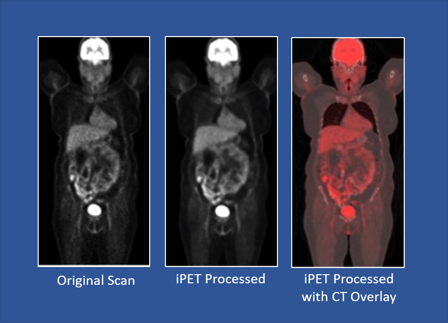
Claritas iPET can be used to enhance Positron Emission Tomography (PET) images with optional simultaneous Magnetic Resonance Imaging (MRI) or Computerized Tomography (CT) scans of the same subject. The image improvement includes noise reduction, sharpening of organ boundaries, and achieving super-resolution. The Claritas iPET algorithm computes the fusion of functional (from PET) and anatomic (from MR or CT) information and is based on Non-Local Means filtering. The goal of the software is to process and visualize the content of DICOM files storing 3D voxel arrays, i.e., a uniformly spaced sequence of slices of a PET scan.
The processing algorithm may also input another 3D voxel array storing the density values obtained by a CT or MRI scan. The PET and CT/MR volumes should at least partially overlap to exploit the additional anatomic information.
How iPET works
- Enhances PET image quality by implementation of an image processing and image fusion algorithm
- The Claritas iPET algorithm computes the fusion of functional (from PET) and anatomic (from MR or CT) information
- The sharpness, style and the detail of the visualization can be controlled by the user
- No new feature is introduced that did not exist in the PET data
- Only existing features are emphasized if they are also supported by the anatomy or suppressed if they are in the noise region and are not supported by the anatomy.
iPET Enhancement Quantified
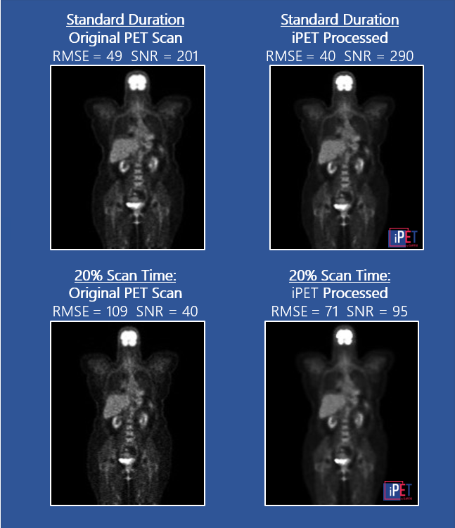
- In enhancing the quality of PET scans, the aim is to increase Signal-to-Noise-Ratio (SNR) and to reduce Root Mean Square Error (RMSE)
- For images captured with low contrast dose and /or short duration scans – iPET improves SNR by 4-5 times and reduces RMSE by 50%
- For images captured as per current standards, iPET improves SNR by up to 20% and reduces RMSE by 10%
- Scan times can be reduced by up to 80% while yielding similar results as that of a full duration scan
Access and Deployment
Seamless Integration into Existing Systems
iPET is designed for easy and seamless integration into your current workflow. With highly configurable and flexible APIs, iPET can be integrated into any PACS system. Integration and deployment is rapid and the Claritas team will provide technical support for smooth integration to optimize image enhancement processing.

Stand-alone Mode
For purposes of research and demonstration, and in the absence of PACS Server, iPET in stand-alone mode runs on the client computer and provides its own 2D/3D graphical user interface.Users may select input DICOM files which are processed by the iPET program running on the client computer.
Data Privacy
Claritas solutions ensure patient privacy is protected and its medical software products are compliant with the Health Insurance Portability and Accountability Act (HIPAA) of the USA, the General Data Protection Regulation (GDPR) of the EU and the Personal Data Protection Act (PDPA) of Singapore.